
 |
 |
|
|---|---|---|
| I & C Technical Guides | ||
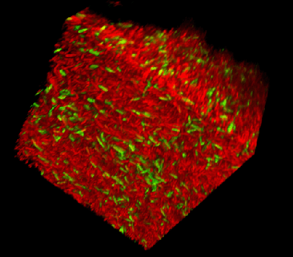
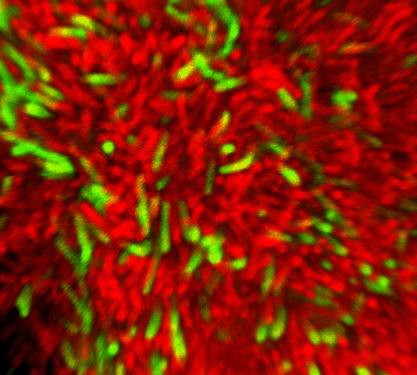
Biofilms are communities of cells attached to a surface. Significant effort is being placed into understanding how they form and the basis for their unique properties, perhaps most notably their resistance to antimicrobials. Imaging technology has and continues to play a very important part in these analyses. Yet, many microbiologists may not be familiar enough with the new technologies to fully maximize their potential. On this site we outline the choices we made to image biofilms, explain what the choices are based on, and list any pitfalls we encountered during imaging and solutions we found. By sharing this information, we hope we can expedite the progress other groups make.
The choice of how to grow a biofilm and how to image should in first instance be driven by the questions that are being asked and realistically, on the technology available and costs. Our overall interest relates to heterogeneity of gene expression in populations, and in this context our goal was to image bacterial biofilms that are growing in the lab in real time with a magnification and resolution that allows individual cells to be identified. Thus, the choices described here are based on this.
 |
 |
|---|
This work and the related publication in the Journal of Microscopy (2009, Volume 235, Pt 2, 128-137) is based on a collaborative effort between University of York microbiologist Marjan van der Woude, based in the Centre for Immunology and Infection (CII) and imaging experts Peter O’Toole and Jo Marrison from the Technology Facility and has been carried out by Matt Lakins (CII).
Modifications to flow cells were carried out by Biosurface Technologies, and other bespoke adaptations by the Department of Biology workshops at University of York, UK .
A detailed hands on course that discusses all aspects of imaging technology is provided by the Technology Facility at the University of York, who also can be approached for contract work. Please go to the Imaging & Cytometry homepage for more information.
| Imaging and Cytometry Laboratory Technology Facility, Department of Biology University of York, PO Box 373 York, YO10 5YW, UK |
Last modified on 3 August 2009 by Jo Marrison |